We do not diagnose disease or recommend a dietary supplement for the treatment of disease. You should share this information with your physician who can determine what nutrition, disease and injury treatment regimen is best for you. You can search this site or the web for topics of interest that I may have written (use Dr Simone and topic).
“We provide truthful information without emotion or influence from the medical establishment, pharmaceutical industry, national organizations, special interest groups or government agencies.” Charles B Simone, M.MS., M.D.
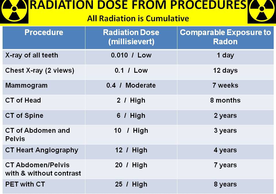
RADIATION DOSE FROM PROCEDURES
https://bit.ly/2p0PLSP
With kind permission from Cancer and Nutrition (Charles B. Simone, M.MS., M.D.)
Lawrenceville, NJ (Dr Charles B Simone) – The electromagnetic radiation spectrum includes gamma rays, X-rays, ultraviolet light, visible light, infrared, microwaves, FM radio waves, AM radio waves, and long radio waves. Electromagnetic radiation with wavelengths longer than the color red (ranging from infrared to radio waves) or shorter than the color violet (ranging from ultraviolet to X-rays and gamma rays) is not visible to the eye. Radiation exposure is another risk factor for developing cancer.
Anne’s History
Anne is 46 and received radiation to her face to treat acne. She also remembers putting her feet in an X-ray machine to see her feet in her new shoes. She has had 5 mammograms and sleeps with an electric blanket and clock radio near her head.
S

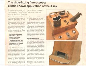
There were more than 10,000 Shoe-fitting Fluoroscopes in the U.S. by the 1940s having 2 or 3 viewing scopes allowing the store clerk, parent and child to see the fit of a shoe on the child’s foot. Children were exposed to High amounts of radiation (1.16 Gy) if they tried on 2 or more pairs of shoes. Burns, stunting of bone and cartilage growth led to the banning of these machines in 1970 in 33 states. How many of you remember this? Source: HemOnc Today May 10, 2008
DIAGNOSTIC RADIATION
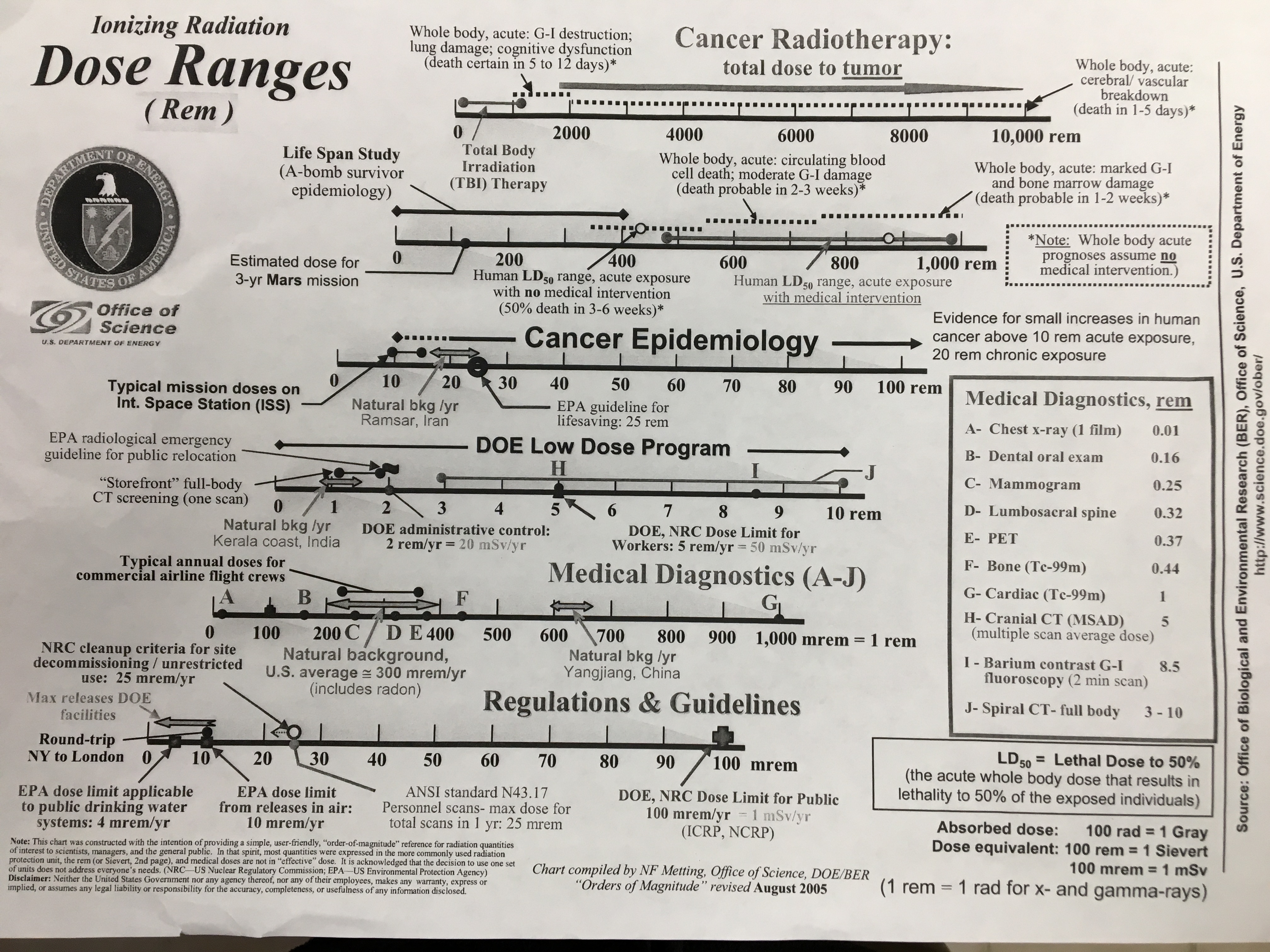
Ionizing radiation causes cancer. Mammograms, CT scans, therapeutic radiation, and other radiation exposures increase the risk for developing cancer.
In fact, about 15,000 people die every year from cancers caused by radiation from CT scans. And children who had a CT scan had a 24% higher risk for developing cancer, with a 16% additional risk for each additional scan. Children who had one CT scan before age 5 had a 35% higher risk for cancer.
Mammogram. The radiation dose from current mammographic two-view examination techniques (1-8 mGy) is extremely damaging to the glandular tissue even though it is less today than before.
The National Academy of Sciences and the National Cancer Institute estimate that for every 100,000 women at age 40 who are screened using mammography, there will be 10 more cancers than are normally seen in that age group due solely to mammograms. The estimate of 10 excess cases is based on the delivery of 1 cGy per mammogram per woman – the amount of radiation delivered by a technically good mammogram unit. However, as more radiation is delivered with older units or poorly calibrated units, this number will be higher based on the higher dose delivered to each patient.
CT Scans. Almost 85 percent of the radiation to which we are exposed in developed countries is from natural sources, but 15 percent is from human-made sources. Of these, about 97 percent is from diagnostic radiology – mainly CT scans.
Children commonly receive an overdose of radiation when they have CT scans. In fact, they receive doses that are at least five times greater than necessary. Radiologists can reduce the dose without compromising image quality but they do not. The number of indications CT scans are used for children has increased dramatically. So if a child today has two or three CT scans of the same area – each giving 2 to 3 cGy per CT scan – over the long term, the risk of cancer is increased significantly.
Do children who hit their heads need a CT scan? Usually not. A careful physical exam is all that is usually necessary. The exceptions are: if the physician suspects a skull fracture, or bleeding in the brain, or the child was in a car crash, or fell off a bike without a helmet, or is unconscious, or has hearing or visual loss.
The lifetime risk of developing cancer attributable to diagnostic X-rays is 1% to 2%, except in Japan where it is 3.2%.5 Table 1 shows the actual number of cancers per year in various countries relative to the number of X-rays given per 1000 people.
Table 1. Number of Cancers per Year from Diagnostic X-rays
Annual X-rays Number
per 1000 people Cancers
Australia 565 431
Canada 892 784
Croatia 903 169
Czech Rep 883 172
Finland 704 50
Germany 1,254 2,049
Japan 1,477 7,587
Kuwait 896 40
Netherlands 600 208
Norway 708 77
Poland 641 291
Sweden 568 162
Switzerland 750 173
United Kingdom 489 700
United States 962 5,695


